Renal Blood Vessels Labeled / Renal Blood Vessels Labeled Renal Artery Doppler Sonographic Tendencies The Hepatic System Is Important Because It Collects Blood From The Intestine And Passes It To The Liver The Centre For : In normal ageing, it drops by around 8ml/min per decade after the age of 30.
Renal Blood Vessels Labeled / Renal Blood Vessels Labeled Renal Artery Doppler Sonographic Tendencies The Hepatic System Is Important Because It Collects Blood From The Intestine And Passes It To The Liver The Centre For : In normal ageing, it drops by around 8ml/min per decade after the age of 30.. Use key choices to identify the blood vessel tunic described. Renal vessels arise at the level of the intervertebral disc between l1 and l2 vertebrae. Berandarenal blood vessels labeled / renal circulation alila medical images : A two pictures of arteries/veins in different locations in. Renal hilum renal pelvis renal sinus (with adipose) major calyx minor calyx renal.
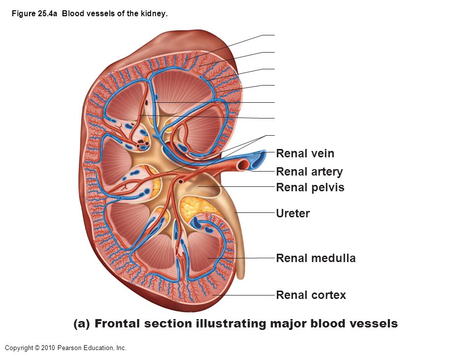
The renal artery enters the hilum of the kidney and divides into a series of smaller vessels. From these arterioles branch the afferent arterioles.each afferent arteriole divides into a capillary network. Blood vessels of the kidney. Renal anatomy (surface anatomy (major blood vessels (celiac: Blood supply of the kidney:
Figure 15 2a The Human Urinary System Ppt Video Online Download from slideplayer.com A part of the blood from the left ventricle is transferred to the kidneys. Compare the anatomy of the sheep kidney to the human kidney. Anatomy of blood vessels review sheet 32 261 microscopic structure of the blood vessels 1. The right renal vein receives tributaries exclusively from the kidney, while the left renal vein receives several tributaries from other organs, including the left. Renal hilum renal pelvis renal sinus (with adipose) major calyx minor calyx renal. Oxygenated blood comes to the kidneys from the right and left renal arteries off the abdominal aorta. Renal blood vessels anatomy the kidneys are highly vascular and thus are equipped with vast and intricate networks of circulation in order to effectively cleanse and modify vast amounts of blood.the hilum permits the entry of the arterial blood flow via the renal artery.the renal artery then branches off creating the interlobular arteries.these. The kidneys' function is dependent on a constant blood supply, so interruptions in the blood flow to the kidneys may result in tissue death and loss of kidney function.
The primary function of large blood vessels (i.e., arteries and veins) is the transport of blood to and from the heart, whereas smaller blood vessels.
The kidneys' function is dependent on a constant blood supply, so interruptions in the blood flow to the kidneys may result in tissue death and loss of kidney function. Because the kidney filters blood, its network of blood vessels is an important component of its structure and function. Anatomy oxygenated blood pumped by the heart passes through the aorta on its way to the kidneys. Blood circulation into and out of the kidneys is highlighted with colored arrows. In normal ageing, it drops by around 8ml/min per decade after the age of 30. Blood vessel changes progressively reduce renal blood flow and gfr: Emerging from the hilum is the renal pelvis, which is formed from the major and minor calyxes in the kidney. This page provides histology support information for blood vessel structure. Its smooth surface decreases resistance to blood flow Does not cover the pathology content. Oxygenated blood comes to the kidneys from the right and left renal arteries off the abdominal aorta. Renal anatomy (surface anatomy, nervous system, vasculature, retroperitoneal space of abdomen (not alot of focus here), posterior abdominal region, viscera of posterior abdominal) Filtered blood leaves the glomerulus via the efferent arteriole, which becomes the interlobular vein.
The interlobar arteries which pass between the renal pyramids, arch around the base of the pyramid as the arcuate. Emerging from the hilum is the renal pelvis, which is formed from the major and minor calyxes in the kidney. Renal anatomy (surface anatomy, nervous system, vasculature, retroperitoneal space of abdomen (not alot of focus here), posterior abdominal region, viscera of posterior abdominal) These give off a series of branches which enter the cortex as interlobular arterioles. Renal blood vessels labeled :
Renal Blood Vessels Labeled Renal Artery Doppler Sonographic Tendencies The Hepatic System Is Important Because It Collects Blood From The Intestine And Passes It To The Liver The Centre For from i0.wp.com Renal blood vessels anatomy the kidneys are highly vascular and thus are equipped with vast and intricate networks of circulation in order to effectively cleanse and modify vast amounts of blood.the hilum permits the entry of the arterial blood flow via the renal artery.the renal artery then branches off creating the interlobular arteries.these. Blood vessels anatomy and physiology anatomy drawing diagram / the process of tubular secretion helps to secrete the urea from the blood to the collecting duct which is finally excreted in form of urine. They ultimately end as afferent arterioles, which transport blood into the renal glomerulus for filtration. A part of the blood from the left ventricle is transferred to the kidneys. Anatomy oxygenated blood pumped by the heart passes through the aorta on its way to the kidneys. The interlobar arteries which pass between the renal pyramids, arch around the base of the pyramid as the arcuate. Renal blood vessels labeled : Filtered blood leaves the glomerulus via the efferent arteriole, which becomes the interlobular vein.
Oxygenated blood enters the kidney from the descending aorta via the renal artery.in the renal hilum, the renal artery divides into segmental arteries, followed by further branching to form interlobar arteries, which pass through the renal columns toward the renal cortex.at the bases of the renal pyramids, the interlobar arteries branch into arcuate arteries, which extend along the arched.
Renal blood vessels anatomy the kidneys are highly vascular and thus are equipped with vast and intricate networks of circulation in order to effectively cleanse and modify vast amounts of blood.the hilum permits the entry of the arterial blood flow via the renal artery.the renal artery then branches off creating the interlobular arteries.these. They ultimately end as afferent arterioles, which transport blood into the renal glomerulus for filtration. These vessels are key elements of kidney function which will be examined shortly during the description of the nephron. Renal vein (vena renalis) the renal vein is an asymmetrically paired vessel that carries the deoxygenated blood from the kidney to the inferior vena cava.both left and right veins run anterior to their corresponding renal arteries. Get ready for your renal blood vessels tests by reviewing key facts, theories, examples, synonyms and definitions with study sets created by students like you. Blood circulation into and out of the kidneys is highlighted with colored arrows. Its smooth surface decreases resistance to blood flow Choose from 500 different sets of kidneys anatomy blood vessels flashcards on quizlet. The renal artery enters the hilum of the kidney and divides into a series of smaller vessels. Anatomy oxygenated blood pumped by the heart passes through the aorta on its way to the kidneys. The renal arteries then progressively branch around the renal pyramids in the following order: Bulky middle tunic contains smooth muscle and elastin 3. Renal anatomy (surface anatomy, nervous system, vasculature, retroperitoneal space of abdomen (not alot of focus here), posterior abdominal region, viscera of posterior abdominal)
A part of the blood from the left ventricle is transferred to the kidneys. The arteries, veins, and nerves that supply the kidney enter and exit at the renal hilum. The renal arteries then progressively branch around the renal pyramids in the following order: Complete the review guide upon completion of the dissection. Blood vessel names and roles are explained in this video, beginning with renal artery and ending with the cortical radiate arteries that serve the glomeruli.
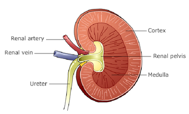
Kidney Structure Biology Notes For Igcse 2014 from biology-igcse.weebly.com Identify the anatomical structures of the kidney. Renal vein (vena renalis) the renal vein is an asymmetrically paired vessel that carries the deoxygenated blood from the kidney to the inferior vena cava.both left and right veins run anterior to their corresponding renal arteries. Renal blood vessels anatomy the kidneys are highly vascular and thus are equipped with vast and intricate networks of circulation in order to effectively cleanse and modify vast amounts of blood.the hilum permits the entry of the arterial blood flow via the renal artery.the renal artery then branches off creating the interlobular arteries.these. The renal arteries then progressively branch around the renal pyramids in the following order: Bloodvessel — the blood vessels are part of the circulatory system and function to transport blood throughout the body. Oxygenated blood enters the kidney from the descending aorta via the renal artery.in the renal hilum, the renal artery divides into segmental arteries, followed by further branching to form interlobar arteries, which pass through the renal columns toward the renal cortex.at the bases of the renal pyramids, the interlobar arteries branch into arcuate arteries, which extend along the arched. This page provides histology support information for blood vessel structure. Get ready for your renal blood vessels tests by reviewing key facts, theories, examples, synonyms and definitions with study sets created by students like you.
Does not cover the pathology content.
In normal ageing, it drops by around 8ml/min per decade after the age of 30. Easy to use and portable, study sets in renal blood vessels are great for studying in the way that works for you, at the time that works for you. Renal hilum renal pelvis renal sinus (with adipose) major calyx minor calyx renal. The renal cortex and medulla contain a complex network of blood vessels. Make sure that you understand the functions of these blood vessels (use your textbook as a resource) renal arteries. Emerging from the hilum is the renal pelvis, which is formed from the major and minor calyxes in the kidney. Oxygenated blood enters the kidney from the descending aorta via the renal artery.in the renal hilum, the renal artery divides into segmental arteries, followed by further branching to form interlobar arteries, which pass through the renal columns toward the renal cortex.at the bases of the renal pyramids, the interlobar arteries branch into arcuate arteries, which extend along the arched. This page provides histology support information for blood vessel structure. The primary function of large blood vessels (i.e., arteries and veins) is the transport of blood to and from the heart, whereas smaller blood vessels. Blood supply of the kidney: Bulky middle tunic contains smooth muscle and elastin 3. Reduced gfr means reduced clearance Anatomy of blood vessels review sheet 32 261 microscopic structure of the blood vessels 1.
Make sure that you understand the functions of these blood vessels (use your textbook as a resource) renal arteries blood vessels labeled. These give off a series of branches which enter the cortex as interlobular arterioles.


0 Komentar